Electromedicine has been around almost as long as we have. The first recorded use of electricity in healing dates back to more than 2,000 years ago when electric eels were used to reduce pain in arthritis sufferers. It wasn’t until the end of the 1700’s when Luigi Galvani, an Italian anatomist, discovered the muscles of dissected frogs twitched whenever they came in contact with two different metals. He concluded the twitching was evidence for the existence of “animal magnetism or electricity,” and was later shown to be erroneous by a contemporary, Volta. However, Galvani was ultimately correct in the existence of an electrical potential in all living cells, just that it was not sufficient to cause the twitching he had observed. Some 40 years later the galvanic principles were so popularly discussed as to become the likely genesis of 18 year old author, Mary Shelley’s novel of Frankenstein.
 It was another hundred years before the little electron would be put to medical use when scientists investigating cathode rays created from free electrons by ionization of the residual air in high voltage discharge tubes invented around 1875 observed their effects. The high voltage Direct Current (DC) of up to 100 kilovolts accelerated the electrons coming from the cathode to a high enough velocity that they created X-rays when they struck the anode or the glass wall of the tube. The German physicist Wilhelm Röntgen was the first to systematically study these effects in 1895 and gave them the name X-Ray. He used them to ‘photograph’ his wife’s hand. Nikola Tesla in his 1897 lecture to the New York Academy of Sciences on methods to generate X-Rays proposed a safer method to generate X-rays and alerted the scientific community to their biological hazard.
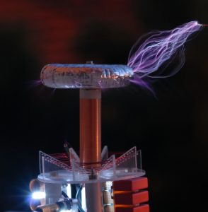
 Most therapeutic applications of electricity at the turn of the previous century were through an inductive connection, rather than a direct connection between the electrical source and the body. This inductive connection involved generating high frequencies and high voltages to create a large electrical field and was generally referred to as Electromedicine. Tesla’s discoveries of using condensers with his coils and a spark gap to create high frequency oscillations of a very high potential, or voltage, were in general therapeutic use by 1900. A French physician and physicist, D’Arsonval, had been using Tesla’s oscillators long before 1900. After receiving several awards from the government of France for his therapeutic use of electrical currents Tesla traveled to Paris in 1892 to confront him. D’Arsonval freely acknowledged his use of Tesla’s oscillators in his work and it was simply the result of French media failing to publish the whole story and wanting to look good in the world’s eye. Dr. D’Arsonval validated the therapeutic benefits of Electromedicine and it became one of the mainstream therapies for treating illness.
Nikola Tesla on November 17, 1898, writing in The Electrical Engineer, Vol. XXVI., No. 550, about therapeutic applications of electricity, reported, “One of the early observed and remarkable features of the high frequency currents, and one which was chiefly of interest to the physician, was their apparent harmlessness which made it possible to pass relatively great amounts of electrical energy through the body of a person without causing pain or serious discomfort. This peculiarity which, together with other mostly unlooked-for properties of these currents I had the honor to bring to the attention of scientific men first in an article in a technical journal in February, 1891, and in subsequent contributions to scientific societies, made it at once evident that these currents would lend themselves particularly to electro-therapeutic uses.”
While X-Rays were being created with extremely high voltage DC, Tesla was creating another source of electron emissions using rare gases and high voltage Alternating Current (AC), resulting in what is commonly known as the neon tube. Tesla understood resonance and how a small amount of energy at the right frequency or vibration could have an additive and sometimes dramatic affect. Building on this principle of resonance and using Tesla’s neon tube, Dr. Royal Raymond Rife in the early 1900’s used radio frequencies that were modulated or pulsed to vary them and then studied their affects in biological samples under an extremely powerful microscope he also invented. Dr. Rife discovered certain frequencies would disrupt the cellular structure of pathogens and even viruses. He used his Rife Rays to treat cancer patients and had a remarkably high success rate.
Tesla freely acknowledged that while he understood how to safely apply these currents to the human body, it “… remained for the physician to investigate the specific actions on the organism and indicate proper methods of treatment…” Unfortunately, physicians speak Latin and engineers speak Greek, and other than Dr. Royal Rife, doctors were unable to explain the specific actions of high frequency currents on the body.
The death-blow to Electromedicine came with The Flexner report of 1910, which was funded heavily by the Rockefeller family who had large investments in the pharmaceutical industry. The report declared frequency based therapies as unscientific and by 1940 Dr. Rife had been discredited and his research and instruments seized by the US government. At about the same time, just prior to his death, Nikola Tesla announced he was about to release a consumer version of vibrational energy medicine device, but it never saw the light of day for many of the same reasons. Dr. Rife’s work of using resonant energies to destroy viruses has since been revitalized and applied by Dr. KT Tsen at Arizona State University in Tempe using coherent laser light to treat blood from patients as it is cycled through a dialysis machine and has successfully demonstrating the ability to safely destroy bacterial microphages.
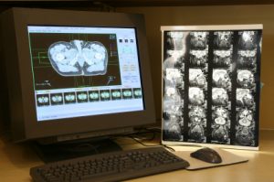
 Using electrons for imaging got a boost in safety in 1950 with the invention of Nuclear Magnetic Resonance Imaging. Nuclear was dropped form the name as patients thought it referred to nuclear bombs during the cold war era, rather than the nucleus of the cells it was imaging. MRI operates by placing the patient in a strong magnetic field, measured in Tesla’s in honor of Nikola Tesla. This magnetic field polarizes the atoms in the body causing them to line up. An RF pulse tuned to the resonant frequency of hydrogen atoms for a given magnetic field strength is then transmitted to a small area of the body. The tuned energy is absorbed by the hydrogen atoms in that area exciting their electrons into a higher orbit around the nucleus of the hydrogen atom. When the short RF pulse is turned off the electrons ‘fall’ back to the normal orbit and release the stored energy which is captured by the MRI receiver and an image is computed. Thus an MRI actually provides an image of the energy in the body, rather than an image of the issues itself, as an X-Ray or CT-Scan does. MRI’s are many times safer than an X-Ray or CT-Scan, as there is significantly less energy transmitted to or through the body.
In 1972, almost two hundred years after Galvani was experimenting with electrical currents applied directly to the body, rather than through inductive means, the first patent was issued for a Transcutaneous Electrical Nerve Stimulator (TENS). These devices provide mild to uncomfortable electric pulses to repeatedly contract and hopefully relax tight muscles. It was also discovered that TENS could overwhelm the Central Nervous System (CNS) and effectively interrupt the pain signals being sent to the brain. TENS have since been used for muscle strengthening and building and most people only receive short term relief with TENS.
Dr. Robert Becker published his findings in 1983 with regard to the regenerative abilities of electricity applied directly to the body. Dr. Becker had observed the reversal of bio-electrical current emanating during regeneration of an amputated appendage in salamanders, as opposed to the diminishing of the bio-electric current during the healing of an amputated appendage in frogs and mice. He theorized that supplanting the bio-electric current in frog and mice with an external DC source that amputated appendages could be regenerated and successfully demonstrated this. Amazing as this sounds, ‘modern’ medicine has generally ignored the dramatic implications of his findings.
Continuing with applications of direct current directly to living tissues, Björn E. W. Nordenström of Sweden proposed a method of treating cancer using his theory of biologically closed electric circuits (BCEC) by inserting an electrode directly into cancerous tissues and causing a current to flow to literally kill the cancer cells. Known as electrochemotherapy (EChT), the AMA rejected his proposals in 1986 and Dr. Nordenström worked instead with China which has seen a marked improvement in cancer treatments using this method.
The original Rife Ray Machine emitted electrons at specific frequencies to kill pathogens in the body. Inductive coupling using radio frequencies is a very inefficient method of transferring electrical energy to a living organism, as it requires tens of thousands of volts and accompanying safety protocols when handling high of voltage to simply raise the cell potential a few thousandths of a volt. SCENAR, which stands for Self Controlled Energy Neuro Adaptive Regulator, and was ostensibly developed for the Russian Space program, applies specific frequencies and waveforms through direct contact to the body like TENS, but at a lower power. This has expanded into a field of Microcurrent Electrotherapy, or MET, which presents very low currents to the body as specific therapeutic frequencies. The use of MET gained popularity starting in 1997 when a chiropractor in Portland, Oregon acquired a used frequency machine from the 1950s and began using it her practice.
One of the accepted uses of Electromedicine that survived the Flexner report is the inductive coupling use of electromagnetic fields to encourage the healing of bone unions that fail to regrow after a break.
Shortwave diathermy works on the principle of using RF energy to warm biological tissues below the surface of the skin and have been sold and used for many years. New devices are disposable battery powered units that use RF energy emitted through a flat antenna which is modulated at a lower therapeutic frequency. Modulating the RF energy has two benefits, one is preventing the build-up of RF energy near the surface of the skin which could cause burns and secondly it introduces Pulsed ElectroMagnetic Fields (PEMF) operating at any one or number of “Rife” frequencies, as most therapeutic frequencies have come to be called. PEMF has been used successfully for over 50 years to aid in the healing of bone fractures and to greatly speed the healing of open sores and rashes on the skin.
 Ionizing foot baths are a newer outgrowth of Electromedicine that have seen growing use. Though the use of the word ionizing is a bit of a misnomer, as the footbaths’ energize the water by passing electrical current between electrodes submerged in the water with the patient’s feet. Discoloration of the water after treatment is purported to be due to toxins removed from the patients, yet study has shown the discoloration is due principally to the oxidation of the metals in the electrodes.
 The availability of low cost consumer microprocessors and industrial microcontrollers has contributed to a proliferation of Electromedicine devices, though most are merely updates of preexisting devices and provide little improvement of previous designs. NuTesla’s instruments are based upon the original resonant energy discoveries of Nikola Tesla represent new and unique applications of these emerging technologies.
